Glaucoma, a leading cause of irreversible blindness, poses a growing global health concern. As the number of cases continues to rise, early detection becomes paramount in preventing vision loss. However, this silent disease often remains undetected until significant damage has occurred, highlighting the need for improved diagnostic methods.
How Doctors Traditionally Diagnose Glaucoma
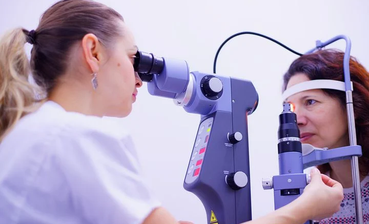
The traditional glaucoma diagnosis relies on a combination of tools and assessments:
- Optic Nerve Exam: Eye doctors examine the optic nerve, located at the back of the eye, for signs of damage characteristic of glaucoma. However, subtle early-stage changes can be difficult to detect.
- Visual Field Test: This test maps a patient’s peripheral vision. Certain patterns of vision loss may indicate glaucoma, but this test can be subjective, with results varying depending on patient effort and concentration.
- Medical History and Risk Factors: Doctors assess factors like age, family history, and other health conditions that may increase glaucoma risk.
Limitations of Traditional Methods
While these methods play a role, they have limitations, particularly in catching early-stage glaucoma:
- Subjectivity: Both optic nerve exams and visual field tests can be subjective, with interpretation varying between examiners.
- Slow Progression: Glaucoma often progresses slowly, meaning subtle, early changes may be missed during routine exams.
The Breakthrough of Optical Coherence Tomography (OCT)
OCT technology overcomes many of the challenges of traditional glaucoma detection:
- Unparalleled Detail: OCT provides high-resolution, cross-sectional images of the eye’s internal structures, revealing the optic nerve and retina with incredible precision.
- Early Detection: OCT can detect subtle changes that may precede traditional signs of glaucoma, offering an opportunity for earlier intervention.
- Objective Measurement: OCT quantifies changes in eye structures, providing doctors with objective data to track the disease’s progression over time.
- Patient Comfort: This quick and painless scan requires no eye drops and is generally well-tolerated.
Why Ganglion Cell Analysis is Key for Glaucoma Detection
Glaucoma causes irreversible damage to the eye’s optic nerve, and ganglion cells are a crucial part of this nerve. They transmit visual information from the eye to the brain. Traditional glaucoma detection methods have limitations, but focusing on ganglion cells offers several advantages.
Firstly, ganglion cells begin to die off before major vision loss occurs, even before noticeable changes in the optic nerve itself. Analyzing these cells offers a chance to detect glaucoma in its earliest stages.
Secondly, Optical Coherence Tomography creates incredibly detailed images of the eye, allowing doctors to measure the thickness of the ganglion cell layer. Thinning of this layer is a major red flag for glaucoma.
Finally, monitoring changes in the ganglion cell layer can help doctors track glaucoma’s progression, evaluating how well treatments are working.
Combining OCT with artificial intelligence makes ganglion cell analysis even more potent for early glaucoma detection. AI algorithms in a platform like Altris AI for OCT for glaucoma analysis detect subtle changes in the ganglion cell layer, meaning even earlier glaucoma detection is possible.
By combining the precision of OCT imaging with AI-powered analysis, we have a powerful new tool to help preserve people’s sight. OCT AI is not the future, it’s the present.
Article edited and fact checked by our editorial team.
References:
- Schuster AK, Erb C, Hoffmann EM, Dietlein T, Pfeiffer N. The Diagnosis and Treatment of Glaucoma. Dtsch Arztebl Int. 2020 Mar 27;117(13):225-234. doi: 10.3238/arztebl.2020.0225. PMID: 32343668; PMCID: PMC7196841.
- Wagner IV, Stewart MW, Dorairaj SK. Updates on the Diagnosis and Management of Glaucoma. Mayo Clin Proc Innov Qual Outcomes. 2022 Nov 16;6(6):618-635. doi: 10.1016/j.mayocpiqo.2022.09.007. PMID: 36405987; PMCID: PMC9673042.
- Chaurasia AK, Greatbatch CJ, Hewitt AW. Diagnostic Accuracy of Artificial Intelligence in Glaucoma Screening and Clinical Practice. J Glaucoma. 2022 May 1;31(5):285-299. doi: 10.1097/IJG.0000000000002015. Epub 2022 Mar 18. PMID: 35302538.