Radiology is a branch of medicine that allows a doctor to see different parts of the body. The process is divided into different procedures, each using a different test which takes pictures of the body parts. Radiography includes tests such as X-ray, MRI, ultrasound, CT scan, and PET scan. You can read a guide to Radiology Information Systems here.
Who are radiologists?
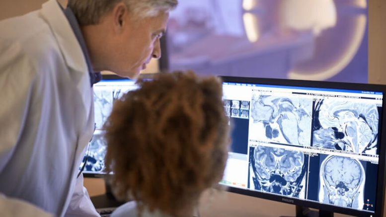
Radiography is an amazingly innovative healthcare field. Radiologists use cutting-edge technologies to examine patients and identify the roots of their conditions, and are a fundamental cog in the incredible engine of therapeutic diagnosis. There are several different radiography jobs currently available:
Radiography assistant
A radiography assistant is a support job that allows radiographers to work even more smoothly and efficiently. Radiography assistants work side by side with diagnostic radiographers and therapeutic radiographers by performing tasks such as processing images, inputting data, and checking equipment. Their salary ranges from £31,000 to £38,000 per year on average.
Diagnostic Radiographer
Diagnostic radiographers use the most advanced radiology technologies to peek inside the patients’ bodies, take images of their insides and help improve the diagnostic process. They are the ones who provide answers to questions like “Is Retrolisthesis the same as Spondylolisthesis“? Their salary ranges from £38,000 to £44,000 per year on average.
Radiography therapist
A radiography therapist or therapeutic radiographer helps cancer patient get better. Radiotherapists use radiotherapy in tandem with other specialists such as oncologists and surgeons to design treatment programmes for cancer patients. Their salary ranges from £31,000 to £38,000 per year on average.
Different types of Radiology
- Diagnostic Radiology
- Interventional radiology
- Radiation Oncology
Diagnostic radiology
Diagnostic radiology is the process of getting scanned images without introducing any instrument inside the body of the patient. In the process, the patients are subjected to quite a low frequency of radiations to create a high definition image of the body part. These radiations involve Radiography (X-rays), Magnetic Resonance Imaging Scan or MRI scan, Computed Tomography Scans or CT, Ultrasound, and Nuclear Medicine Scans.
The process can be used to diagnose a number of problems, such as broken bones, blood clots, and heart conditions; moreover, even gastrointestinal conditions can be diagnosed with this method. This method is effective to diagnose severe diseases such as breast cancer and is less invasive – usually requiring just some ultrasound gel to be applied on the section of the body that is examined. In addition to identifying issues, diagnostic radiology can be used by physicians to understand how well your body is responding to current treatment.
Interventional Radiology
This imaging technique utilizes various image-forming processes, such as ultrasound, x-ray fluoroscopy, computed tomography or magnetic resonance imaging. An interventional radiologist is able to treat and diagnose numerous diseases by inserting tiny instruments into the body via arteries.
The images formed from this process are used by doctors to locate the exact tissue which needs to be treated and can be further used to monitor the expanse of a disease. Through this method, the specialists use minimal space-invasive techniques to diagnose problems through an image.
Radiation Oncology
Radiation Oncology is an effective mode of treatment of cancer which makes use of radiation therapy or radiations, for treating cancer. It is also an effective way of getting an image of body parts. Studies claim that approximately 40% of the total patients who have cancer across the world are cured with this therapy.
What is radiation therapy?
Radiation therapy can be used to kill the cancer cells present in the body. It can prevent their growth, multiplication and can even stop them from spreading in different parts of the body. Do healthy cells also get affected by the radiation?
Yes! However, the cancerous cells get more affected by radiation. Thus, it is mandatory to consult with a professional before you step on to any conclusion. Therefore, physicians prefer this method above than any other method. Types of radiation used for the treatment and getting the image of the body parts includes:
- X-rays
- Gamma rays
- Electron beams
Benefits of Radiology
Now that we know what Radiology is let’s find out how it can be beneficial for us. Following are some reasons why most physicians and doctors use it to prevent the spread and ill-effects of certain diseases:
- With radiology, doctors can skip the surgery intended to diagnose an ailment.
- It helps you understand whether you would still need the surgery.
- It assists in making a diagnosis and helps doctors come up with a further plan for the management of most body conditions.
- Interventional radiology, the advanced form of diagnosis, involves treatment also. With this method, the risk is lesser, and so is the recovery time!
- Doctors use radiology to diagnose diseases such as breast cancer through mammography, and therefore, suppresses the mortality rate.
- Therapies involving radiology can even help treat cancer.
Disadvantages of Radiology
While this technology confers enormous benefits, it has some disadvantages too. The electronic images obtained via Radiology are very easy to modify, and therefore, every image obtained from radiological scanners comes with a doubt on its reliability. Therefore, this doubt has questioned the validity of the images for the use of experiments.
Is Radiology Risky?
Unavoidably, during a radiological exam, patients are exposed to a certain amount of radiations. To minimize the risks for health, however, radiations are kept at a minimum and are focused only on the area that needs to be examined.X-rays are so safe, in fact, that even children can undergo the procedure.
The only exception are pregnant women, for whom radiographies are usually contraindicated because the fetus is particularly vulnerable during the early development phase. Radiographies are usually avoided during pregnancy, unless they’re absolutely necessary (such as when facing a serious medical emergency). In these rare occasions, additional safety precautions are taken such as covering the patient’s body with a special protective apron.
On the other hand, a radiologist might be exposed to radiations on a daily basis, so even small amounts might eventually become a threat. To prevent this from becoming an issue, they take appropriate safety measures, such as using special protections.
Article edited and fact checked by our editorial team.
References:
- Linton O. What is radiology? Acad Radiol. 2007 Nov;14(11):1433. doi: 10.1016/j.acra.2007.07.025.
- Evidence-Based Radiology Working Group. Evidence-based radiology: a new approach to the practice of radiology. Radiology. 2001 Sep;220(3):566-75. doi: 10.1148/radiol.2203001465.
- “Radiology — Diagnostic Specialty Description”. American Medical Association. Retrieved from: https://freida.ama-assn.org/specialty/radiology-diagnostic
- Kreel L. Medical imaging. Postgrad Med J. 1991 Apr;67(786):334-46. doi: 10.1136/pgmj.67.786.334.